Embark on an intriguing journey into the chambers of the heart crossword, where the intricate workings of this vital organ are laid bare. This comprehensive guide delves into the anatomy, physiology, and potential health concerns associated with the heart’s chambers, providing a thorough understanding of their significance in maintaining cardiovascular health.
From the right atrium to the left ventricle, we’ll explore the unique functions of each chamber and trace the pathway of blood flow through the heart. Along the way, we’ll uncover the role of valves in regulating blood flow, examine the cardiac cycle, and discuss common heart murmurs and chamber abnormalities.
Chambers of the Heart: Chambers Of The Heart Crossword

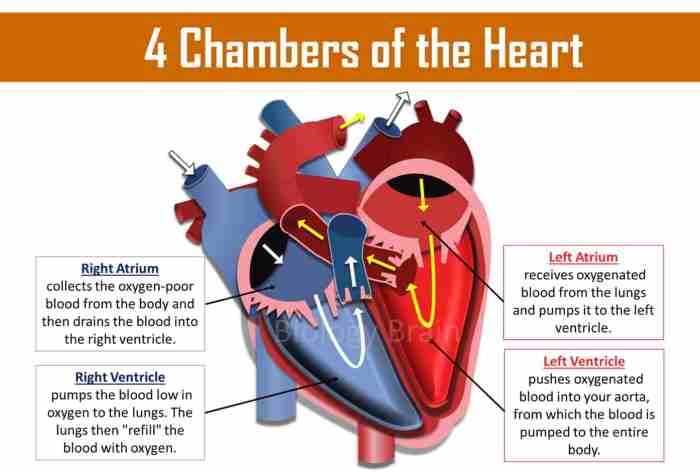
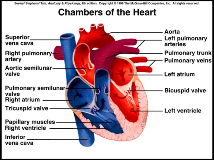
The heart is a muscular organ that pumps blood throughout the body. It has four chambers: two atria (upper chambers) and two ventricles (lower chambers).
The right atrium receives deoxygenated blood from the body and pumps it to the right ventricle. The right ventricle then pumps the blood to the lungs, where it picks up oxygen.
The left atrium receives oxygenated blood from the lungs and pumps it to the left ventricle. The left ventricle then pumps the blood to the body.
| Chamber | Function |
|---|---|
| Right atrium | Receives deoxygenated blood from the body |
| Right ventricle | Pumps blood to the lungs |
| Left atrium | Receives oxygenated blood from the lungs |
| Left ventricle | Pumps blood to the body |
Blood Flow through the Chambers

The heart is a remarkable organ responsible for pumping oxygenated blood throughout the body. Understanding the pathway of blood flow through its chambers is crucial for comprehending the heart’s vital role in maintaining circulation.
Blood flow through the heart follows a specific sequence, ensuring that oxygen-rich blood is delivered to the body while deoxygenated blood is returned to the lungs for reoxygenation.
Deoxygenated Blood Flow
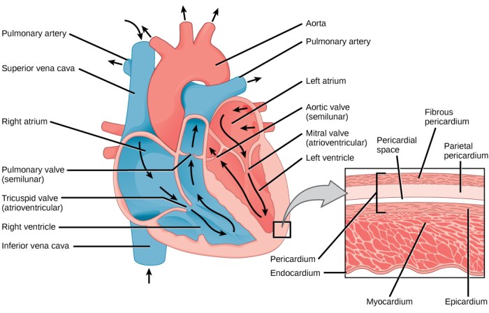
The journey begins with deoxygenated blood entering the heart through two large veins, the superior vena cava and the inferior vena cava. These veins carry blood from the body back to the heart.
- Right Atrium:The deoxygenated blood first enters the right atrium, the heart’s upper right chamber. From here, it flows into the right ventricle, the lower right chamber.
- Right Ventricle:The right ventricle contracts, pumping the deoxygenated blood into the pulmonary artery, which carries it to the lungs for reoxygenation.
Oxygenated Blood Flow
Once oxygenated in the lungs, blood returns to the heart via the pulmonary veins. These veins carry oxygen-rich blood to the left atrium, the heart’s upper left chamber.
- Left Atrium:From the left atrium, blood flows into the left ventricle, the lower left chamber.
- Left Ventricle:The left ventricle contracts, pumping the oxygenated blood into the aorta, the largest artery in the body. The aorta distributes the oxygen-rich blood to various organs and tissues throughout the body.
Role of Valves, Chambers of the heart crossword
Throughout the blood flow pathway, valves play a crucial role in regulating blood flow and preventing backflow.
- Atrioventricular Valves:Located between the atria and ventricles, these valves (tricuspid valve on the right and mitral valve on the left) prevent blood from flowing back into the atria during ventricular contraction.
- Semilunar Valves:Found at the exits of the ventricles, these valves (pulmonary valve on the right and aortic valve on the left) prevent blood from flowing back into the ventricles during ventricular relaxation.
Cardiac Cycle and Chamber Function

The cardiac cycle is the rhythmic contraction and relaxation of the heart, which pumps blood throughout the body. It consists of two main phases: systole and diastole.
During systole, the heart contracts and pumps blood out of the chambers. The atria contract first, pushing blood into the ventricles. Then, the ventricles contract, pumping blood out of the heart and into the arteries.
During diastole, the heart relaxes and fills with blood. The atria fill first, and then the ventricles fill. The cycle then repeats.
Role of the Chambers in Pumping Blood
The atria are the receiving chambers of the heart, and the ventricles are the pumping chambers. The atria contract to push blood into the ventricles, and the ventricles contract to pump blood out of the heart.
Chaining your brain with a chambers of the heart crossword? Take a breather and check out this interesting tidbit: a baseball rolls off a 0.70 m platform, how far will it roll? Now, back to your crossword puzzle, where the right atrium and ventricle pump blood to the lungs, while the left atrium and ventricle pump blood to the body.
Keep solving!
The right atrium receives blood from the body and pumps it into the right ventricle. The right ventricle then pumps the blood to the lungs, where it is oxygenated.
The left atrium receives oxygenated blood from the lungs and pumps it into the left ventricle. The left ventricle then pumps the blood out to the body.
Heart Murmurs and Chamber Abnormalities
Heart murmurs are abnormal sounds produced by the heart during its pumping action. They can be caused by various factors, including chamber abnormalities.
Chamber abnormalities, such as septal defects (holes in the heart’s walls) or valve abnormalities, can disrupt the normal blood flow through the heart. This can create turbulence and abnormal vibrations, which can be detected as heart murmurs.
Common Heart Murmurs Associated with Chamber Abnormalities
- Atrial septal defect (ASD):This is a hole in the wall between the right and left atria. It can cause a murmur that sounds like a “whooshing” noise.
- Ventricular septal defect (VSD):This is a hole in the wall between the right and left ventricles. It can cause a murmur that sounds like a “blowing” noise.
- Patent ductus arteriosus (PDA):This is a connection between the aorta and the pulmonary artery that normally closes after birth. If it remains open, it can cause a murmur that sounds like a “machinery” noise.
Diagnostic Techniques Used to Detect Chamber Abnormalities
Chamber abnormalities can be detected using various diagnostic techniques, including:
- Echocardiography:This is an ultrasound imaging technique that uses sound waves to create images of the heart. It can show the structure and function of the heart, including any abnormalities.
- Cardiac catheterization:This is a procedure in which a thin tube (catheter) is inserted into the heart through an artery or vein. It can measure pressures and oxygen levels in the heart, and can also be used to inject contrast dye to visualize the heart’s chambers.
- Electrocardiography (ECG):This is a test that records the electrical activity of the heart. It can show the heart’s rhythm and rate, and can also help detect abnormalities in the heart’s electrical system.
Congenital Heart Defects Involving Chambers
Congenital heart defects involving the chambers arise during fetal development and affect the structure and function of the heart’s chambers. These defects can range in severity, from mild abnormalities to life-threatening conditions.
Types of Congenital Heart Defects Involving Chambers
- Atrial Septal Defect (ASD):A hole in the atrial septum, the wall separating the right and left atria, allowing blood to flow abnormally between the chambers.
- Ventricular Septal Defect (VSD):A hole in the ventricular septum, the wall separating the right and left ventricles, leading to abnormal blood flow between the chambers.
- Tetralogy of Fallot:A combination of four heart defects, including a VSD, pulmonary valve stenosis (narrowing of the valve leading to the lungs), an overriding aorta (aorta positioned over both ventricles), and right ventricular hypertrophy (enlargement of the right ventricle).
- Transposition of the Great Arteries:A defect where the aorta arises from the right ventricle and the pulmonary artery from the left ventricle, reversing the normal blood flow pattern.
Symptoms and Diagnosis
Symptoms of congenital heart defects involving chambers can vary depending on the type and severity of the defect. Common symptoms include:
- Cyanosis (bluish skin, lips, or nail beds due to low oxygen levels)
- Shortness of breath or rapid breathing
- Fatigue or exercise intolerance
- Chest pain or palpitations
Diagnosis typically involves a combination of physical examination, medical history, echocardiography (ultrasound of the heart), and other imaging tests.
Treatment Options
Treatment options for congenital heart defects involving chambers vary depending on the specific defect and its severity. Treatment may include:
- Medication:To manage symptoms and prevent complications
- Cardiac catheterization:A minimally invasive procedure to close or repair defects
- Open-heart surgery:To correct the defect and restore normal heart function
Chamber Enlargement and Dysfunction

Chamber enlargement, also known as cardiomegaly, occurs when the heart’s chambers become abnormally large. This condition can result from various causes, including hypertension, valvular heart disease, and cardiomyopathy. Cardiomegaly can lead to heart failure if left untreated.Chamber dysfunction refers to the impaired ability of the heart’s chambers to pump blood effectively.
This can be caused by conditions such as heart failure, arrhythmias, and valvular heart disease. Chamber dysfunction can significantly affect heart function, leading to symptoms such as shortness of breath, fatigue, and chest pain.
Diagnostic Tests for Chamber Size and Function
Several diagnostic tests can be used to assess chamber size and function. These include:
Echocardiography
An ultrasound of the heart that provides images of the heart’s chambers and valves, allowing doctors to assess their size and function.
Cardiac MRI
A magnetic resonance imaging scan of the heart that provides detailed images of the heart’s chambers and valves, enabling doctors to evaluate their size and function.
Chest X-ray
A plain X-ray of the chest that can show the size and shape of the heart’s chambers.
Chamber-Specific Diseases and Conditions
The heart’s chambers can be affected by a variety of diseases and conditions. Some of these conditions primarily affect specific chambers, while others can affect multiple chambers.
Atrial Fibrillation
Atrial fibrillation is a heart rhythm disorder that causes the atria to contract irregularly and rapidly. This can lead to a variety of symptoms, including palpitations, shortness of breath, fatigue, and lightheadedness. Atrial fibrillation is diagnosed with an electrocardiogram (ECG) and is typically treated with medication or ablation therapy.
Right Ventricular Hypertrophy
Right ventricular hypertrophy is a condition in which the right ventricle becomes enlarged and thickened. This can be caused by a variety of conditions, including pulmonary hypertension, tricuspid valve regurgitation, and congenital heart defects. Right ventricular hypertrophy can lead to a variety of symptoms, including shortness of breath, fatigue, and swelling in the legs and ankles.
It is diagnosed with an echocardiogram and is typically treated with medication or surgery.
Left Ventricular Failure
Left ventricular failure is a condition in which the left ventricle is unable to pump enough blood to meet the body’s needs. This can be caused by a variety of conditions, including coronary artery disease, heart attack, and cardiomyopathy. Left ventricular failure can lead to a variety of symptoms, including shortness of breath, fatigue, and swelling in the legs and ankles.
It is diagnosed with an echocardiogram and is typically treated with medication, lifestyle changes, and sometimes surgery.
Top FAQs
What are the four chambers of the heart?
The four chambers of the heart are the right atrium, right ventricle, left atrium, and left ventricle.
What is the function of the right atrium?
The right atrium receives deoxygenated blood from the body and pumps it into the right ventricle.
What is the cardiac cycle?
The cardiac cycle is the sequence of events that occur during one heartbeat, including systole (contraction) and diastole (relaxation).